Synthetic Platelets Put the Brakes on Blood Loss
In animals, they cut bleeding time in half, but use in humans is still far off
By Ed EdelsonHealthDay Reporter
WEDNESDAY, Dec. 16 (HealthDay News) -- Hoping to improve on nature, researchers have built and tested synthetic versions of the blood-clotting cells called platelets, to be used in trauma or other cases where blood just won't stop flowing.
dblclick('xxlA');
"We start by making a core, with material that is used in degradable stitches, which dissolve in the body," said Erin B. Lavik, a professor of biomedical engineering at Case Western Reserve University, and lead author of a report published Dec. 16 in Science Translational Medicine. "Then we attach a polymer that is soluble in water and is used in the pharmaceutical industry. Then we attach a molecule that interacts with activated platelets and helps them clot more quickly."
The hope is that the artificial platelets can replace or augment the activity of the currently used clotting medication, known as factor VIIa, Lavik explained.
Factor VIIa is a protein that plays a central role in blood clotting. A genetically engineered version of the protein is now available for medical use. It was introduced for use in people with hemophilia, a genetic condition in which normal clotting does not occur, and it is being increasingly used against uncontrollable hemorrhage.
But factor VIIa must be kept in refrigerated form and has a short shelf life, Lavik said. And it cannot be used for head or spinal cord injuries, for fear of complications.
"The reason we developed this synthetic platelet is that it is stable at all temperatures," Lavik said. "It is a fine powder that can be administered intravenously. The faster you can control bleeding, the better the outcome."
In animal tests, injured rats given injections of the artificial platelets stopped bleeding in half the time of those that went untreated. Rats that got injections 20 seconds after an injury stopped bleeding in 23 percent less time than untreated rats.
"We also did head-to-head comparisons with factor VIIa," Lavik said. "When the artificial platelets were introduced, bleeding was reduced even more." The artificial platelets induced clotting 25 percent faster than factor VIIa, the report said.
However, a long series of tests lie ahead before the artificial platelets can enter routine medical practice, Lavik said. "The next step would be an animal model that most closely mimics human injury," she said. "We have to move up to larger animals. Pigs are most commonly used."
Financial help is also needed. "We have applied to federal and non-federal groups for funding," Lavik said. She is hoping for support from a pharmaceutical company because "ultimately, you have to think about making it commercially viable."
"We have just started having those conversations," Lavik said. "Now that we have published this paper, we hope we can generate some interest."
A critical point is convincing the U.S. Food and Drug Administration that artificial platelets would have a useful medical application, Lavik said. Convincing the FDA would start with data from future animal studies. "Assuming that it replicates what we have seen so far, then we would talk with the FDA," she said. "There is no use estimating our chance of success until we see that data and talk with them."
"Anything new that would be safe to use with someone who has ongoing hemorrhage would be useful in a trauma center," added Dr. Michael Craun, trauma medical director at Scott and White Memorial Hospital in Temple, Texas. "We really have problems now with people who have major injuries."
Another expert agreed. "Any compound or device that can stem hemorrhage in patients can be helpful if the risk-benefit ratio is favorable," said Dr. Brian Harbrecht, director of trauma at the University of Louisville.
But he and Craun also stressed the early nature of the work.
"A lot more investigation needs to go into this particular product to see if it is clinically applicable or not," Harbrecht said. "That requires years and years of more precise work."
The new report described "preliminary experiments with rats only," Craun added, and there are questions about safety, cost and technology still to be answered
Thursday, December 17, 2009

Wednesday, December 16, 2009
52 Facts About Blood Donation
1. More than 4.5 million people need blood transfusions each year in the U.S. and Canada.
2. 43,000 pints: amount of donated blood used each day in the U.S. and Canada.
3. Someone needs blood every two seconds.
4. 37% of the U.S. population is eligible to donate blood – less than 10% do annually**.
5. About 1 in 7 people entering a hospital need blood.
6. One pint of blood can save up to three lives.
7. Healthy people who are at least 17 years old (16 with parental consent), and at least 110 pounds may donate whole blood every 56 days. Females receive 53% of blood transfusions; males receive 47%.
8. 94% of blood donors are registered voters.
9. In 1901, Dr. Karl Landsteiner first identified the major human blood groups: A, B, AB and O.
10. People with O- blood are universal donors of red blood cells.
11. People with AB+ blood are universal recipients of red blood cells, and universal donors of plasma.
12. One unit of whole blood can be separated into several components, including red blood cells, plasma, and platelets.
13. Red blood cells carry oxygen to the body's organs and tissues, and live for about 120 days in the circulatory system.
14. Platelets promote blood clotting and give those with leukemia and other cancers a chance to live.
15. Plasma is a pale yellow mixture of water, proteins and salts.
16. Plasma, which is 90% water, makes up 55% of blood volume.
17. Healthy bone marrow makes a constant supply of red cells, plasma and platelets.
18. Blood or plasma that comes from people who have been paid for it cannot be used for human transfusion.
19. Granulocytes, a type of white blood cell, roll along blood vessel walls in search of bacteria to engulf and destroy.
20. White cells are the body's primary defense against infection.
21. Apheresis is a special kind of blood donation that allows a donor to give specific blood components, such as platelets or red blood cells.
22. 42 days: how long most donated red blood cells can be stored.
23. Five days: how long most donated platelets can be stored.
24. One year: how long frozen plasma can be stored.
25. Much of today's medical care depends on a steady supply of blood from healthy donors.
26. 2.7 pints: the average whole blood and red blood cell transfusion.*
27. Children being treated for cancer, premature infants and children having heart surgery may receive blood and platelets during their treatments.
28. Anemic patients may need blood transfusions to increase their red blood cell levels.
29. Cancer, transplant and trauma patients, and patients undergoing open-heart surgery may require platelet transfusions to survive.
30. Sickle cell disease is an inherited disease that affects more than 80,000 people in the U.S., 98% of whom are of African descent.
31. Many patients with severe sickle cell disease receive blood transfusions every month.
32. Over 10 tests are performed on each unit of donated blood.
33. 17% of non-donors cite "never thought about it" as the main reason for not giving blood, while 15% say they're too busy.
34. The #1 reason blood donors say they give is because they "want to help others."
35. Blood centers often run short of types O and B red blood cells.
36. There is no substitute for human blood.
37. If all blood donors gave three times a year, blood shortages would be a rare event (The current average is about two).
38. 46.5 gallons: amount of blood you could donate if you begin at age 17 and donate every 56 days until you are 79 years old.
39. There are four easy steps to donate blood: medical history, a quick physical, donation and snacks.
40. The actual blood donation takes less than 15 minutes. The entire process – from the time you sign in until the time you leave – usually takes under an hour.
41. After donating blood, you replace the fluid in hours and the red blood cells within four weeks. It takes eight weeks to restore the iron lost after donating.
42. You cannot get AIDS or any other infectious disease by donating blood.
43. 10 pints: the amount of blood in the body of an average adult.
44. One unit of whole blood is roughly the equivalent of one pint.
45. Blood makes up about 7% of your body's weight.
46. Newborn babies have about one cup of blood in their bodies.
47. Giving blood will not decrease your strength.
48. Any company, community organization, place of worship or individual may contact their local community blood center to host a blood drive.
49. Roughly half of all blood donations across the U.S. are collected at blood drives.
50. People who donate blood are volunteers and are not paid for their donation.
51. 500,000 Americans donated blood in the days following the events of September 11.
52. Blood donation. It's about an hour of your time. It's About Life!
2. 43,000 pints: amount of donated blood used each day in the U.S. and Canada.
3. Someone needs blood every two seconds.
4. 37% of the U.S. population is eligible to donate blood – less than 10% do annually**.
5. About 1 in 7 people entering a hospital need blood.
6. One pint of blood can save up to three lives.
7. Healthy people who are at least 17 years old (16 with parental consent), and at least 110 pounds may donate whole blood every 56 days. Females receive 53% of blood transfusions; males receive 47%.
8. 94% of blood donors are registered voters.
9. In 1901, Dr. Karl Landsteiner first identified the major human blood groups: A, B, AB and O.
10. People with O- blood are universal donors of red blood cells.
11. People with AB+ blood are universal recipients of red blood cells, and universal donors of plasma.
12. One unit of whole blood can be separated into several components, including red blood cells, plasma, and platelets.
13. Red blood cells carry oxygen to the body's organs and tissues, and live for about 120 days in the circulatory system.
14. Platelets promote blood clotting and give those with leukemia and other cancers a chance to live.
15. Plasma is a pale yellow mixture of water, proteins and salts.
16. Plasma, which is 90% water, makes up 55% of blood volume.
17. Healthy bone marrow makes a constant supply of red cells, plasma and platelets.
18. Blood or plasma that comes from people who have been paid for it cannot be used for human transfusion.
19. Granulocytes, a type of white blood cell, roll along blood vessel walls in search of bacteria to engulf and destroy.
20. White cells are the body's primary defense against infection.
21. Apheresis is a special kind of blood donation that allows a donor to give specific blood components, such as platelets or red blood cells.
22. 42 days: how long most donated red blood cells can be stored.
23. Five days: how long most donated platelets can be stored.
24. One year: how long frozen plasma can be stored.
25. Much of today's medical care depends on a steady supply of blood from healthy donors.
26. 2.7 pints: the average whole blood and red blood cell transfusion.*
27. Children being treated for cancer, premature infants and children having heart surgery may receive blood and platelets during their treatments.
28. Anemic patients may need blood transfusions to increase their red blood cell levels.
29. Cancer, transplant and trauma patients, and patients undergoing open-heart surgery may require platelet transfusions to survive.
30. Sickle cell disease is an inherited disease that affects more than 80,000 people in the U.S., 98% of whom are of African descent.
31. Many patients with severe sickle cell disease receive blood transfusions every month.
32. Over 10 tests are performed on each unit of donated blood.
33. 17% of non-donors cite "never thought about it" as the main reason for not giving blood, while 15% say they're too busy.
34. The #1 reason blood donors say they give is because they "want to help others."
35. Blood centers often run short of types O and B red blood cells.
36. There is no substitute for human blood.
37. If all blood donors gave three times a year, blood shortages would be a rare event (The current average is about two).
38. 46.5 gallons: amount of blood you could donate if you begin at age 17 and donate every 56 days until you are 79 years old.
39. There are four easy steps to donate blood: medical history, a quick physical, donation and snacks.
40. The actual blood donation takes less than 15 minutes. The entire process – from the time you sign in until the time you leave – usually takes under an hour.
41. After donating blood, you replace the fluid in hours and the red blood cells within four weeks. It takes eight weeks to restore the iron lost after donating.
42. You cannot get AIDS or any other infectious disease by donating blood.
43. 10 pints: the amount of blood in the body of an average adult.
44. One unit of whole blood is roughly the equivalent of one pint.
45. Blood makes up about 7% of your body's weight.
46. Newborn babies have about one cup of blood in their bodies.
47. Giving blood will not decrease your strength.
48. Any company, community organization, place of worship or individual may contact their local community blood center to host a blood drive.
49. Roughly half of all blood donations across the U.S. are collected at blood drives.
50. People who donate blood are volunteers and are not paid for their donation.
51. 500,000 Americans donated blood in the days following the events of September 11.
52. Blood donation. It's about an hour of your time. It's About Life!
Tuesday, December 15, 2009
Fake blood 2.0?
Posted by Bob Grant
[Entry posted at 14th December 2009 09:14 PM GMT]
View comment(1) Comment on this news story
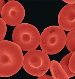
Newly created synthetic particles that mimic red blood cells may one day carry drug molecules and/or oxygen through bloodstreams, according to researchers writing in this week's issue of the Proceedings of the National Academy of Sciences (PNAS). What's more, the team of scientists in Michigan and California say the particles could also be used to improve the resolution of magnetic resonance imaging.
The synthetic red blood cellsthat Mitragotri and his team developedImage: Nishit Doshi"It's a very nice paper and very exciting work," Krishnendo Roy, a biomedical engineer at the University of Texas at Austin who wasn't involved with the study, told The Scientist. "The beauty of their method is its simplicity." University of California, Santa Barbara, chemical engineer Samir Mitragotri led the team of scientists and told The Scientist that the blood cell-like particles could evolve into useful tools in the clinic. "What we got very excited about was making a structure with synthetic materials that begins to mimic a natural object," said Mitragotri. "If we can bridge the gap [between synthetic materials and living cells] it will open up tremendous opportunities for synthetic materials." Mitragotri said that he and his team tested the ability of the particles to carry oxygen, finding that they had a "comparable" oxygen-carrying capacity to actual red blood cells. He added that it may be possible in the future to link therapeutic agents destined for the vascular system, such as heparin, to the particles so that they can be easily distributed throughout the blood. The artificial blood cells, with attached iron oxide nanoparticles, could also one day improve MRI resolution by serving as contrast agents that provide a different imaging signal compared to the surrounding tissue, Mitragotri said. Mitragotri and his colleagues created the artificial red blood cells by first making tiny spheres out of a biodegradable polymer called poly(lactic acid-co-glycolide) (PLGA). They then exposed the spheres to isopropanol, which collapsed them into the discoid shape characteristic of red blood cells. The researchers then layered proteins -- either albumin or hemoglobin -- onto the doughnut-shaped disks, cross-linked the proteins to give them extra strength and stability, and finally dissolved away the PLGA template to leave only a strong but flexible shell of proteins in the shape and size (about 7 microns in diameter) of a red blood cell. Mitragotri and his team then tested the ability of the artificial cells to behave like real blood cells, passing them through glass capillary tubes that were narrower than the diameter of the particles and testing their oxygen-carrying capacity. They showed that the particles could carry about 90 percent of the oxygen real red blood cells can carry. They also showed that a drug-mimicking molecule could easily be loaded into and off of the artificial blood cells. "They conclusively demonstrated some stuff concerning oxygen-carrying capacity and the potential for drug release," Patrick Doyle, a chemical engineer at the Massachusetts Institute of Technology who was not involved with the study, told The Scientist. But years of continued testing lie between Mitragotri's synthetic red blood cells and clinical application. Several questions, including how long the particles will remain in circulation, how the immune system will react to the synthetic blood cells, and how efficiently they transport oxygen, remain to be answered. Mitragotri said that his lab plans on answering these questions by studying the particles in model organisms, research that is set to begin soon. "Whether this is applicable in an in vivo setting," said Roy, "we won't know that for 3, 4, or 5 years." "I don't think these [clinical applications] are far off ideas, but you have to go through all the usual regulatory hurdles," said Doyle, noting that the synthetic cells might also be used to study how cellular aberrations, such as tumor cells, behave in the body. "Ultimately they can also be model systems, by which you can understand disease states of cells," he added.
Friday, October 23, 2009
Oswald Hope Robertson
Jump to: navigation, search
Oswald Hope Robertson (2 June 1886 – 23 March 1966) was an English-born medical scientist who pioneered the idea of blood banks in the "blood depots" he established in 1917 during service in France with the US Army Medical Corps.
Robertson was born in Woolwich in south-east London, but at the age of one-and-a-half he emigrated with his parents to California, settling in the San Joaquin Valley. He attended local schools in Dinuba, then graduated from the Polytechnic High School in San Francisco.
His initial plan to study basic biology was changed by a meeting with an American medical student while on holiday in Germany. After attending some lectures on anatomy, he decided to study medicine, being admitted to the University of California in 1906. He later studied at Harvard Medical School, the Massachusetts General Hospital and the Rockefeller Institute for Medical Research, but had to cut short his studies during World War I when he was called to join medical teams in France. Here he experimented with preserving human blood cells for use in blood transfusions, and became recognised as the inventor of the blood bank.
[edit] Commemoration
Jump to: navigation, search
Oswald Hope Robertson (2 June 1886 – 23 March 1966) was an English-born medical scientist who pioneered the idea of blood banks in the "blood depots" he established in 1917 during service in France with the US Army Medical Corps.
Robertson was born in Woolwich in south-east London, but at the age of one-and-a-half he emigrated with his parents to California, settling in the San Joaquin Valley. He attended local schools in Dinuba, then graduated from the Polytechnic High School in San Francisco.
His initial plan to study basic biology was changed by a meeting with an American medical student while on holiday in Germany. After attending some lectures on anatomy, he decided to study medicine, being admitted to the University of California in 1906. He later studied at Harvard Medical School, the Massachusetts General Hospital and the Rockefeller Institute for Medical Research, but had to cut short his studies during World War I when he was called to join medical teams in France. Here he experimented with preserving human blood cells for use in blood transfusions, and became recognised as the inventor of the blood bank.
[edit] Commemoration
Thursday, April 23, 2009
Conslusion
As already seen in the discussion part, the hypothesis which expressed there is no relationship between activation/inhibition of the platelets and externalization the phosphatidylserine would be, based on the results, ruled out.
Another hypothesis which expresses the relation ship is still on the table. It is not possible to declare:
-Activation of the platelets by Thrombin causes externalization of phosphatidylserine on the surface of the platelets.
-In this experiment the same results could be not applied to ADP as the test was performed for different concentrations of ADP but satisfactory results were not reached. Changing method in this regard in suggested.
-Inhibition of the platelets by Aspirin causes internalization of phosphatidylserine on the surface of the platelets. This statement has been approved by comparing the results of controls with and with out Aspirin.
In addition, having seen the papers, we would realize that COHEN Zoë and colleagues conducted a research in 2004 and their find out declare that if platelets become activated, they would express phosphatidylserine on the outer leaflet of the plasma membrane. They have working with ADP as they say ADP-induced PS exposure. They wanted to realise the role of Caspase in this process as well, while we were not. It can be a suggestion for further works as well to consider different enzymes in this process. Their findings could be useful to prove the activatory role of ADP as well.
Finally, the characteristics of Annexin V have been approved again. The property which they bind with negatively charged phospholipid on the surface of the cells, although it is not the main statement of this experiment but this viable facts could be used to be applied in other experiments as well.
6.1. Suggestions for further works
It should be realised how Thrombin/ADP or Aspirin apply their changes on the platelets. If we were working on cells which have nuclei the possibility of effect on the nuclei could be mentioned. But the platelets which are circulating in blood are not same cell. Therefore changing the expression of a gene which is highly possible for physiological reactions is ruled out. The other and simple way which comes to mind is acting by the aid of a second messenger. Further studies should be based on understanding the mechanism of this effect.
Repeating this experiment with different activators is suggested as well, in particular due to unclear results from ADP.
Further experiments should investigate about the role of Caspase and try to find out the mechanism of inhibition and activation with more details.
As stated about the receptors of ADP and Thrombin. The mechanism of action of stimulation (activation or inhibition) should be considered to find more relationship between the factor of change and response.
Another hypothesis which expresses the relation ship is still on the table. It is not possible to declare:
-Activation of the platelets by Thrombin causes externalization of phosphatidylserine on the surface of the platelets.
-In this experiment the same results could be not applied to ADP as the test was performed for different concentrations of ADP but satisfactory results were not reached. Changing method in this regard in suggested.
-Inhibition of the platelets by Aspirin causes internalization of phosphatidylserine on the surface of the platelets. This statement has been approved by comparing the results of controls with and with out Aspirin.
In addition, having seen the papers, we would realize that COHEN Zoë and colleagues conducted a research in 2004 and their find out declare that if platelets become activated, they would express phosphatidylserine on the outer leaflet of the plasma membrane. They have working with ADP as they say ADP-induced PS exposure. They wanted to realise the role of Caspase in this process as well, while we were not. It can be a suggestion for further works as well to consider different enzymes in this process. Their findings could be useful to prove the activatory role of ADP as well.
Finally, the characteristics of Annexin V have been approved again. The property which they bind with negatively charged phospholipid on the surface of the cells, although it is not the main statement of this experiment but this viable facts could be used to be applied in other experiments as well.
6.1. Suggestions for further works
It should be realised how Thrombin/ADP or Aspirin apply their changes on the platelets. If we were working on cells which have nuclei the possibility of effect on the nuclei could be mentioned. But the platelets which are circulating in blood are not same cell. Therefore changing the expression of a gene which is highly possible for physiological reactions is ruled out. The other and simple way which comes to mind is acting by the aid of a second messenger. Further studies should be based on understanding the mechanism of this effect.
Repeating this experiment with different activators is suggested as well, in particular due to unclear results from ADP.
Further experiments should investigate about the role of Caspase and try to find out the mechanism of inhibition and activation with more details.
As stated about the receptors of ADP and Thrombin. The mechanism of action of stimulation (activation or inhibition) should be considered to find more relationship between the factor of change and response.
Discussion
The hypothesis of this project as stated before is mainly about finding a relationship between activators of platelets (Thrombin and ADP) and Inhibitor of palettes (Aspirin) on externalisation the phosphatidylserine on the outer membrane of the platelets which would bind to Annexin V labelled with fluorescent. Therefore by the end of running the experiment for five times the data should be analysed to find logic relation ship between aforementioned parameters. By comparing the result of flowcytometry, few groups of analysis are reached. After ward in a separate graph they are compared. In all charts Y error bars represent Standard Deviations.
5.1. Thrombin and Aspirin
5.1.1. The first chart will demonstrate the externalisation of the Thrombin in different concentrations. It is with out Aspirin .This chart would reveal that what is the effect of Thrombin in externalisation of Phosphatidylserine with out Aspirin.
Graph 5.1.1.1. Externalising of Phosphatidylserine in different concentrations of Thrombin with out Aspirin.
The first graphs is indicating that , adding Thrombin on its own could rise up externalising phosphatidylserine on the outer membrane of the platelets. This is while Aspirin had not been added to the samples and Alcohol (ethanol) was used instead of Aspirin. As the chart suggests the general trend is increasing.
5.1.2. The next stage is to evaluate the role of Aspirin. The first eight tubes are with out Aspirin (Solution A had been added).The mean of fluorescent intensity for different concentration of Thrombin would be me calculated. For tubes from 8 to 16, solution B had been added which was including Aspirin. Then the mean of Fluorescent intensity for different concentration of Thrombin for these tubes would be taken as well. They should be compared with each other. The result comes below.
Chart 5.1.2.1. Compare the effect of Aspirin on different concentrations of Thrombin.
The concentration of each tube has been mentioned. The added Aspirin per each tube was 2µL. As the graph suggests, in most cases the ADP causes decreasing in externalisation of Thrombin although the change is not significant( P<0.05) however in this regard in a research conducted by Conehn Zoë and colleagues in 2004, revealed that when the plateletes become inhibited they express less phosphatidylserine on the outer membrane. This matches with the achievements of our experiment.
5.2. ADP and Aspirin
Before doing this experiment another method was used in order to get proper data for ADP. But as results were not satisfactory therefore only the double amount of final concentration of ADP is used in new method. In previous method the range of concentration of ADP was similar to Thrombin in current method and highest concentration of ADP was 10µL while in current method the concentration of ADP is 20 µL. Perhaps running this method will not reach a visible result about ADP. The role of ADP in activating the platelets has already been proved (Jianguo Jin et al, 1998) .The fact that activating by ADP cause measurable change is not something which could be found by this experiment. Maybe by applying different methods, better results are reached. Therefore the first chart (like Thrombin) will be excluded.
5.2.1. This stage is to evaluate the effect of Aspirin on platelets which have already been activated by ADP. The tube 8 is include 20µL ADP with out Aspirin (Solution A had been added) and tube 16 is include 20µL ADP with Aspirin. In five times of running the experiment the mean of fluorescent intensity of tubes 8 and 16 would be me calculated and the results are compared with each other. The concentration of added Aspirin as mentioned in method is 0.01 % V/V. The important factor in this regard is the amount of Aspirin used for each tube.
Chart 5.2.2.1. Aspirin has effect on platelets which had already been activated by ADP.
By this chart it is not quite clear to recognise what is the effect of Aspirin of the specimen which have already been activated with ADP. May be there are some interactions between the function of ADP and Aspirin in platelets cell membrane which can cause a visible change. But apart from specimen 4 which has a high SD (as the Y-error bars shows); generally it is logic to say that Aspirin cause internalisation the phosphatidylserine. We should not forget that we are looking for effect of Aspirin (either internalising or externalising). If platelets have already been activated with different activators although they may cause overlap, but the most important impact should not be forgotten. The next chart will reveals more valuable information in this regard.
Additionally, as stated before Aspirin Induces Apoptosis through -Release of Cytochrome c from Mitochondria (Katja C Zimmermann et al, 2000) and the inhibition of Proteasome Function (Priyanka Dikshit et al, 2006). It is logic and true. When a cell is inhibited for along time and is not in use, the final destination is not something apart from apoptosis. Although the main cause of apoptosis is not what was mentioned, but inhibition a cell.
5.3. Control and Aspirin
In order to prove the hypothesis, the sole effect of Aspirin on platelets which have not been activated by any factor would be evaluated as well. This is a compare between controls of samples which have been added Alcohol (from solution A) and the control of samples which have been added Aspirin (from solution B). Chart comes below manifest this compare.
Chart 5.2.2.1. Compare Controls with and with out Aspirin.
Here a significant change is observed (P<0.01). href="http://www.ncbi.nlm.nih.gov/sites/entrez?Db=pubmed&Cmd=Search&Term=%22Casta%C3%B1o%20E%22%5BAuthor%5D&itool=EntrezSystem2.PEntrez.Pubmed.Pubmed_ResultsPanel.Pubmed_RVAbstract">Castaño E and colleagues in 1999 announced that Aspirin increases phosphatidylserine externalization when they were analysing HT-29 colon carcinoma cells .In this paper is said that Aspirin induces cell death and caspase-dependent phosphatidylserine externalization. Based on their paper one of two halls marks of apoptosis is: increase in phosphatidylserine externalization. : It does not correspond with other findings which discuss about inhibitory role of Aspirin. As mentioned before, when platelets become activated they express more phosphatidylserine on the outer leaflet, therefore when they become inhibited (for example by Aspirin) they logically must express less phosphatidylserine while bases on the aforementioned paper was not. But Pamela L in her book demonstrate that unlike observation about colorectal cell lines , if the method of measuring is based on Annexin V binding to phosphatidylserine, there was not dose dependent apoptotic increase .
5.1. Thrombin and Aspirin
5.1.1. The first chart will demonstrate the externalisation of the Thrombin in different concentrations. It is with out Aspirin .This chart would reveal that what is the effect of Thrombin in externalisation of Phosphatidylserine with out Aspirin.
Graph 5.1.1.1. Externalising of Phosphatidylserine in different concentrations of Thrombin with out Aspirin.
The first graphs is indicating that , adding Thrombin on its own could rise up externalising phosphatidylserine on the outer membrane of the platelets. This is while Aspirin had not been added to the samples and Alcohol (ethanol) was used instead of Aspirin. As the chart suggests the general trend is increasing.
5.1.2. The next stage is to evaluate the role of Aspirin. The first eight tubes are with out Aspirin (Solution A had been added).The mean of fluorescent intensity for different concentration of Thrombin would be me calculated. For tubes from 8 to 16, solution B had been added which was including Aspirin. Then the mean of Fluorescent intensity for different concentration of Thrombin for these tubes would be taken as well. They should be compared with each other. The result comes below.
Chart 5.1.2.1. Compare the effect of Aspirin on different concentrations of Thrombin.
The concentration of each tube has been mentioned. The added Aspirin per each tube was 2µL. As the graph suggests, in most cases the ADP causes decreasing in externalisation of Thrombin although the change is not significant( P<0.05) however in this regard in a research conducted by Conehn Zoë and colleagues in 2004, revealed that when the plateletes become inhibited they express less phosphatidylserine on the outer membrane. This matches with the achievements of our experiment.
5.2. ADP and Aspirin
Before doing this experiment another method was used in order to get proper data for ADP. But as results were not satisfactory therefore only the double amount of final concentration of ADP is used in new method. In previous method the range of concentration of ADP was similar to Thrombin in current method and highest concentration of ADP was 10µL while in current method the concentration of ADP is 20 µL. Perhaps running this method will not reach a visible result about ADP. The role of ADP in activating the platelets has already been proved (Jianguo Jin et al, 1998) .The fact that activating by ADP cause measurable change is not something which could be found by this experiment. Maybe by applying different methods, better results are reached. Therefore the first chart (like Thrombin) will be excluded.
5.2.1. This stage is to evaluate the effect of Aspirin on platelets which have already been activated by ADP. The tube 8 is include 20µL ADP with out Aspirin (Solution A had been added) and tube 16 is include 20µL ADP with Aspirin. In five times of running the experiment the mean of fluorescent intensity of tubes 8 and 16 would be me calculated and the results are compared with each other. The concentration of added Aspirin as mentioned in method is 0.01 % V/V. The important factor in this regard is the amount of Aspirin used for each tube.
Chart 5.2.2.1. Aspirin has effect on platelets which had already been activated by ADP.
By this chart it is not quite clear to recognise what is the effect of Aspirin of the specimen which have already been activated with ADP. May be there are some interactions between the function of ADP and Aspirin in platelets cell membrane which can cause a visible change. But apart from specimen 4 which has a high SD (as the Y-error bars shows); generally it is logic to say that Aspirin cause internalisation the phosphatidylserine. We should not forget that we are looking for effect of Aspirin (either internalising or externalising). If platelets have already been activated with different activators although they may cause overlap, but the most important impact should not be forgotten. The next chart will reveals more valuable information in this regard.
Additionally, as stated before Aspirin Induces Apoptosis through -Release of Cytochrome c from Mitochondria (Katja C Zimmermann et al, 2000) and the inhibition of Proteasome Function (Priyanka Dikshit et al, 2006). It is logic and true. When a cell is inhibited for along time and is not in use, the final destination is not something apart from apoptosis. Although the main cause of apoptosis is not what was mentioned, but inhibition a cell.
5.3. Control and Aspirin
In order to prove the hypothesis, the sole effect of Aspirin on platelets which have not been activated by any factor would be evaluated as well. This is a compare between controls of samples which have been added Alcohol (from solution A) and the control of samples which have been added Aspirin (from solution B). Chart comes below manifest this compare.
Chart 5.2.2.1. Compare Controls with and with out Aspirin.
Here a significant change is observed (P<0.01). href="http://www.ncbi.nlm.nih.gov/sites/entrez?Db=pubmed&Cmd=Search&Term=%22Casta%C3%B1o%20E%22%5BAuthor%5D&itool=EntrezSystem2.PEntrez.Pubmed.Pubmed_ResultsPanel.Pubmed_RVAbstract">Castaño E and colleagues in 1999 announced that Aspirin increases phosphatidylserine externalization when they were analysing HT-29 colon carcinoma cells .In this paper is said that Aspirin induces cell death and caspase-dependent phosphatidylserine externalization. Based on their paper one of two halls marks of apoptosis is: increase in phosphatidylserine externalization. : It does not correspond with other findings which discuss about inhibitory role of Aspirin. As mentioned before, when platelets become activated they express more phosphatidylserine on the outer leaflet, therefore when they become inhibited (for example by Aspirin) they logically must express less phosphatidylserine while bases on the aforementioned paper was not. But Pamela L in her book demonstrate that unlike observation about colorectal cell lines , if the method of measuring is based on Annexin V binding to phosphatidylserine, there was not dose dependent apoptotic increase .
4. Experimental results
The results of flowcytometry of each tube come below. The experiment was performed for five times. Hence it is expected to see the five series of sixteen results.
In each group of results two diagrams and one table are seen. Details of each result come below (Guide line of the flowcytometry machine).
-Upper diagram
FSC Forward Scatter: Cell size
SSC Side Scatter: Granularity
-Lower diagram
X scatter: Fluorescent intensity emitted is proportional to the quantity of binding sites for the fluorescent compound on the cell/particle. The unit is MESF which is Molecules of Equivalent Soluble Fluorochrome. In this experiment the geometric mean of fluorescent is measured.
Y scatter: Events count
-Table
Geometric mean of all events
Geometric mean of M1 region (1% of the events)
FL2-H: 585/42 nm, yellow-green colour
Mean: Average X-axis channel number of linear value for events in the quadrant
Geometric Mean: average of the logarithm of the X-axis channel number or linear value for events in the quadrant expressed anti-log.
Picture 4.1.1. Flowcytometry machine.
BD FCSCalibur two laser channel.
4.1. Diagrams and table from flowcytometry.
Results of flowcytometry for each sample come next.
The results of flowcytometry of each tube come below. The experiment was performed for five times. Hence it is expected to see the five series of sixteen results.
In each group of results two diagrams and one table are seen. Details of each result come below (Guide line of the flowcytometry machine).
-Upper diagram
FSC Forward Scatter: Cell size
SSC Side Scatter: Granularity
-Lower diagram
X scatter: Fluorescent intensity emitted is proportional to the quantity of binding sites for the fluorescent compound on the cell/particle. The unit is MESF which is Molecules of Equivalent Soluble Fluorochrome. In this experiment the geometric mean of fluorescent is measured.
Y scatter: Events count
-Table
Geometric mean of all events
Geometric mean of M1 region (1% of the events)
FL2-H: 585/42 nm, yellow-green colour
Mean: Average X-axis channel number of linear value for events in the quadrant
Geometric Mean: average of the logarithm of the X-axis channel number or linear value for events in the quadrant expressed anti-log.
Picture 4.1.1. Flowcytometry machine.
BD FCSCalibur two laser channel.
4.1. Diagrams and table from flowcytometry.
Results of flowcytometry for each sample come next.
M&M
3. Materials & Methods
3.1 Materials
Apart from ADP, Thrombin and Aspirin which have been explained before, further substances are required as well which would be described below.
3.1.1 HEPES buffer
HEPES buffer is a suitable alternative general-purpose zwitterionic buffer. HEPES buffer does not bind to Mg +2, Ca +2, Mn +2 and Cu +2. HEPES buffer is one from ten important biological buffers.
-HEPES buffer with CaCl2 is used in this experiment .It is a zwitterionic buffer does not bind Ca +2 in plasma. HEPES buffer can exhibit toxicity if the concentration is greater than 40 mM .20 mM HEPES is the most satisfactory concentration. HEPES buffer can maintain pH. That is why it is more used in cell culture.
HEPES used in this experiment is consisting of:
-10 mM NaCl, 10mM HEPES (4-(2-hydroxyethyl)-1-piperazineethanesulfonic acid)
,2.7 mM CaCl2 ,5 mM Glucose ,0.5 mg/mL BSA ,Distilled water up to 500mL ,Few drops of NaOH to reach the pH to 7.4 (Lepe-Zuniga JL et al , 1987)
3.1.2. Fixative solution
Fixative solution used in this experiment is formaldehyde also known as methanal, CH2O MW=30.03, which is a very good stabiliser and fixative in biological studies. Usually it is used in 4% w/v concentration. It does limit oxidation and polymerisation.
3.2. Method
3.2.1. General description
The idea if planning this method is based on similar study conducted by Jef L and colleagues in 2006.
In a nut shell the over view of all done in the method of this experiment is:
1-Preparing HEPES buffer
2-Pareparting PRP: Platelets Rich Plasma (Centrifuging in 200 g for 10 minutes)
3-Prepraing series of dilution of Thrombin and ADP.
4-Adding Alcohol (Ethanol) [tubes 1 to 8] and adding Aspirin [tubes 9 to 16]
5- Taking 10µL from indicated tubes +90µL fixative (formaldehyde)
6-Taineg 1ml from above and mixing with 1 ml distilled water.
7-Flowcytometry to plot a histogram and gather the data.
More details comes bellow.
3.2.2 Platelet isolation
First stage is platelets isolation.10 mL blood was collected in 2 mL anticoagulant ACD (anticoagulant citrate dextrose solution) (2.5% sodium citrate, 1.5% citric acid, and 2% glucose).The donor should not take Aspirin within last few days. Then the specimens are centrifuged for 10 minutes in 2000g to reach the Platelet Reach Plasma (PRP). Follow by order HEPES buffer is prepared to use in next stages as the main buffer of this experiment.
3.2.3 Solution A, Solution B
Two separate solutions called solution A and B are prepared as follow:
-Solution A: Solution A is consist of: 2mL buffer + 50 µL PRP + 2µL Alcohol (Ethanol) Concentration of Ethanol = 0.01 % (0.00009746) V/V
-Solution B: Solution B is consist of: 2mL buffer + 50 µL PRP + 2µL Aspirin
Concentration of Aspirin=0.01 % (0.00009746) V/V
3.2.4 Preparing preliminary concentration of Thrombin and ADP
A set of 16 tubes are required. The following concentration of Thrombin and ADP are made. By the end of this stage the concentration of Thrombin and ADP content of each tube will be as the following table.
Table 3.2.4.1. Preliminary concentrations of Thrombin and ADP.
Tube 1
10 µL buffer
Tube 2
10 µL buffer
Tube 3
0.10 Unit/mL Thrombin
Tube 4
0.30 Unit/mL Thrombin
Tube 5
1.00 Unit/mL Thrombin
Tube 6
3.00 Unit/mL Thrombin
Tube 7
10.00 Unit/mL Thrombin
Tube 8
200µM ADP
Tube 9
10µL buffer
Tube 10
10µL buffer
Tube 11
0.10 Unit/mL Thrombin
Tube 12
0.30 Unit/mL Thrombin
Tube 13
1.00 Unit/mL Thrombin
Tube 14
3.00 Unit/mL Thrombin
Tube 15
10.00 Unit/mL Thrombin
Tube 16
200µM ADP
By the end of this stage 16 tubes are ready. Now extra 16 tubes are required to carry on the experiment to prepare the final concentrations of Thrombin and ADP.
3.2.5. Preparing final concentrations of Thrombin and ADP
In these series of tubes we need to take 10 µL from the previous tubes and mix with 90 µL of solution A for tubes 1 to 8 and solution B for tubes 9 to 16. Hence we will see the new set of tubes in which the concentration of thrombin and ADP will become 10 fold less. By the end of this stage in new set of tubes the concentration of Thrombin or ADP and content of each tube will be as the following table.
Table 3.2.5.1. Final concentrations of Thrombin and ADP.
Tube 1
10mL buffer
Tube 2
10mL buffer
Tube 3
0.01 Unit/mL Thrombin
Tube 4
0.03 Unit/mL Thrombin
Tube 5
0.10 Unit/mL Thrombin
Tube 6
0.30 Unit/mL Thrombin
Tube 7
1.00 Unit/mL Thrombin
Tube 8
20µM ADP
Tube 9
10mL buffer
Tube 10
10mL buffer
Tube 11
0.01 Unit/mL Thrombin
Tube 12
0.03 Unit/mL Thrombin
Tube 13
0.10 Unit/mL Thrombin
Tube 14
0.30 Unit/mL Thrombin
Tube 15
1.00 Unit/mL Thrombin
Tube 16
20µM ADP
After this stage we need to wait for ten minutes for each tube .Then Annexin V will be added.
3.2.6. Adding Annexin V and preparing for flowcytometry
At this stage 5µL Annexin V is added to each tube. Annexin V is labelled with fluorescent substance (florin). Then after 50µL of each tube is taken and mixed with 100 µL fixative (formaldehyde) in another tube. We can now leave the tubes for a couple of hours. It is very important to do the flowcytometry at the same day. Although they are fixed with fixative but they should not be kept for more than some hours. Before final stage, 1 mL of each tube is taken and mixed with distilled water in another series of tube. This is to prepare for flowcytometry.
3.2.7. Flowcytometry
The final stage is flowcytometry. The flowcytometry machine will be set as described above and the results would be gathered. The results of Flowcytometry come in next part.
3.1 Materials
Apart from ADP, Thrombin and Aspirin which have been explained before, further substances are required as well which would be described below.
3.1.1 HEPES buffer
HEPES buffer is a suitable alternative general-purpose zwitterionic buffer. HEPES buffer does not bind to Mg +2, Ca +2, Mn +2 and Cu +2. HEPES buffer is one from ten important biological buffers.
-HEPES buffer with CaCl2 is used in this experiment .It is a zwitterionic buffer does not bind Ca +2 in plasma. HEPES buffer can exhibit toxicity if the concentration is greater than 40 mM .20 mM HEPES is the most satisfactory concentration. HEPES buffer can maintain pH. That is why it is more used in cell culture.
HEPES used in this experiment is consisting of:
-10 mM NaCl, 10mM HEPES (4-(2-hydroxyethyl)-1-piperazineethanesulfonic acid)
,2.7 mM CaCl2 ,5 mM Glucose ,0.5 mg/mL BSA ,Distilled water up to 500mL ,Few drops of NaOH to reach the pH to 7.4 (Lepe-Zuniga JL et al , 1987)
3.1.2. Fixative solution
Fixative solution used in this experiment is formaldehyde also known as methanal, CH2O MW=30.03, which is a very good stabiliser and fixative in biological studies. Usually it is used in 4% w/v concentration. It does limit oxidation and polymerisation.
3.2. Method
3.2.1. General description
The idea if planning this method is based on similar study conducted by Jef L and colleagues in 2006.
In a nut shell the over view of all done in the method of this experiment is:
1-Preparing HEPES buffer
2-Pareparting PRP: Platelets Rich Plasma (Centrifuging in 200 g for 10 minutes)
3-Prepraing series of dilution of Thrombin and ADP.
4-Adding Alcohol (Ethanol) [tubes 1 to 8] and adding Aspirin [tubes 9 to 16]
5- Taking 10µL from indicated tubes +90µL fixative (formaldehyde)
6-Taineg 1ml from above and mixing with 1 ml distilled water.
7-Flowcytometry to plot a histogram and gather the data.
More details comes bellow.
3.2.2 Platelet isolation
First stage is platelets isolation.10 mL blood was collected in 2 mL anticoagulant ACD (anticoagulant citrate dextrose solution) (2.5% sodium citrate, 1.5% citric acid, and 2% glucose).The donor should not take Aspirin within last few days. Then the specimens are centrifuged for 10 minutes in 2000g to reach the Platelet Reach Plasma (PRP). Follow by order HEPES buffer is prepared to use in next stages as the main buffer of this experiment.
3.2.3 Solution A, Solution B
Two separate solutions called solution A and B are prepared as follow:
-Solution A: Solution A is consist of: 2mL buffer + 50 µL PRP + 2µL Alcohol (Ethanol) Concentration of Ethanol = 0.01 % (0.00009746) V/V
-Solution B: Solution B is consist of: 2mL buffer + 50 µL PRP + 2µL Aspirin
Concentration of Aspirin=0.01 % (0.00009746) V/V
3.2.4 Preparing preliminary concentration of Thrombin and ADP
A set of 16 tubes are required. The following concentration of Thrombin and ADP are made. By the end of this stage the concentration of Thrombin and ADP content of each tube will be as the following table.
Table 3.2.4.1. Preliminary concentrations of Thrombin and ADP.
Tube 1
10 µL buffer
Tube 2
10 µL buffer
Tube 3
0.10 Unit/mL Thrombin
Tube 4
0.30 Unit/mL Thrombin
Tube 5
1.00 Unit/mL Thrombin
Tube 6
3.00 Unit/mL Thrombin
Tube 7
10.00 Unit/mL Thrombin
Tube 8
200µM ADP
Tube 9
10µL buffer
Tube 10
10µL buffer
Tube 11
0.10 Unit/mL Thrombin
Tube 12
0.30 Unit/mL Thrombin
Tube 13
1.00 Unit/mL Thrombin
Tube 14
3.00 Unit/mL Thrombin
Tube 15
10.00 Unit/mL Thrombin
Tube 16
200µM ADP
By the end of this stage 16 tubes are ready. Now extra 16 tubes are required to carry on the experiment to prepare the final concentrations of Thrombin and ADP.
3.2.5. Preparing final concentrations of Thrombin and ADP
In these series of tubes we need to take 10 µL from the previous tubes and mix with 90 µL of solution A for tubes 1 to 8 and solution B for tubes 9 to 16. Hence we will see the new set of tubes in which the concentration of thrombin and ADP will become 10 fold less. By the end of this stage in new set of tubes the concentration of Thrombin or ADP and content of each tube will be as the following table.
Table 3.2.5.1. Final concentrations of Thrombin and ADP.
Tube 1
10mL buffer
Tube 2
10mL buffer
Tube 3
0.01 Unit/mL Thrombin
Tube 4
0.03 Unit/mL Thrombin
Tube 5
0.10 Unit/mL Thrombin
Tube 6
0.30 Unit/mL Thrombin
Tube 7
1.00 Unit/mL Thrombin
Tube 8
20µM ADP
Tube 9
10mL buffer
Tube 10
10mL buffer
Tube 11
0.01 Unit/mL Thrombin
Tube 12
0.03 Unit/mL Thrombin
Tube 13
0.10 Unit/mL Thrombin
Tube 14
0.30 Unit/mL Thrombin
Tube 15
1.00 Unit/mL Thrombin
Tube 16
20µM ADP
After this stage we need to wait for ten minutes for each tube .Then Annexin V will be added.
3.2.6. Adding Annexin V and preparing for flowcytometry
At this stage 5µL Annexin V is added to each tube. Annexin V is labelled with fluorescent substance (florin). Then after 50µL of each tube is taken and mixed with 100 µL fixative (formaldehyde) in another tube. We can now leave the tubes for a couple of hours. It is very important to do the flowcytometry at the same day. Although they are fixed with fixative but they should not be kept for more than some hours. Before final stage, 1 mL of each tube is taken and mixed with distilled water in another series of tube. This is to prepare for flowcytometry.
3.2.7. Flowcytometry
The final stage is flowcytometry. The flowcytometry machine will be set as described above and the results would be gathered. The results of Flowcytometry come in next part.
.1. Annexin V (Endonexin II, Placental anticoagulant protein I, vascular anticoagulant-alpha, Lipocortin V, Placental protein 4; PP4, Anchorin CII) (Bohn H et al, 1979 ; Ernest Beutler et al, 2001 ;Reutelingsperger CP ,2001 ;Hendrikus H et al , 2005). Annexin V (Gene map locus 4q26-q28), a member of the Annexin family with an unknown function up to now , is a cellular protein which is known as a phosphatidylserine-binding protein (Ernest Beutler et al , 2001) . Annexin family has over 160 proteins. They all share the property of Ca 2+ dependent binding to negatively charged phospholipid surfaces (Gerke V et al, 2002). At late of 70s Annexin V for the first time was purified from an EDTA extract of human placenta (Bohn H et al, 1979; Hendrikus H et al, 2005). After ward, a protein (Fig. 2.1.1.) was discovered independently in blood vessels which was named vascular anticoagulant protein Alpha (VAC-Alpha) which inhibits blood coagulation (Reutelingsperger CP, 1985). The mechanism of anticoagulation is based on the high-affinity binding to Phospholipids (Fiedler K et al, 1995). This is the property which makes Annexin V quite effective in inhibiting the prothrombinase complex (Reutelingsperger CP, 1988; Homburg CH et al, 1995). In vitro, Annexin V is defined as a Phospholipid binding protein with a high affinity for PS (phosphatidylserine) (D’Arceuil H et al, 2000). One of the important functions of Annexin V is forming a shield around negatively-charged phospholipid molecules (Dumont EA et al, 2000; Dreier R et al, 1998). This formation of Annexin V shield would block the entry of phospholipid molecules into coagulation clotting reactions (Glaser M et al, 2003). An increased quantity of phospholipid molecules on cell membranes which speed up coagulation reactions is seen in the absence of the shield(Koopman G et al , 1994 ; Vermes I et al , 1995 ). Annexin V binds quite tightly to anionic phospholipids (Catherine .R et al, 1992). There are evidences suggesting Annexin V interacts with phospholipid vesicles (Hendrikus H et al, 2005). Annexin V can form a shield around phospholipid molecules which blocks their entry into coagulation (clotting) reactions (Ernest Beutler et al, 2001). The formation of this shield is disrupted by the abnormal antibodies in case of anti phospholipid antibody syndrome (Hayes MJ, 2004). In the absence of the shield, there is an increased quantity of phospholipid molecules on cell membranes (Gerke V et al, 2002). These molecules speed up coagulation reactions and cause the abnormal blood clotting which is characteristic of the anti phospholipid antibody syndrome (Ernest Beutler et al, 2001; Huber R et al , 1992).
Figure 2.1.1.X-ray analysis on Annexin V. Tertiary
Structure of Annexin V is revealed (Consists of 319
amino acids).Green spheres are indicating Ca 2+
2.2. Phosphatidylserine - C13H24NO10P (MW: 385.304)
Phosphatidylserine (Fig. 2.2.1.) is a phospholipid component essential to the functioning of all the cells of the body which is mainly known as a molecule which enables brain cells to metabolise glucose (Vermes I et al, 1995). It is mainly located entirely on the inner layer of the plasma membrane. Phosphatidylserine is mainly known as effective treatment for Alzheimer's disease . Phosphatidylserine is usually on the cytosolic side, of cell membranes (Catherine .R et al, 1992; Calderon F et al, 2008). By a flipase enzyme Phosphatidylserine become exposed on the surface when a cell undergoes apoptotic cell death (Boersma et al, 2003; Belhocine T et al, 2002). The normal distribution of Phosphatidylserine is altered during platelet activation and cellular apoptosis (Fadok VA et al, 1992) Phosphatidylserine modulates the activity of several enzymes involved in cellular signalling. Annexins (particularly Annexin V) bind to and polymerize through protein-protein interactions on membrane patches which are expressing phosphatidylserine. Annexin V specifically tends to bind to phosphatidylserine (Calderon F et al, 2008). As stated before Phosphatidylserine is almost exclusively located on the cytoplasmic side of the plasma membrane and after stimulation, Phosphatidylserine becomes exposed on the outer side of plasma membrane at a proportion depending on the type and the nature of the stimuli (Cohen Zoë et al , 2004 ;Kenis H et al ,2004).
Figure 2.2.1.Chemical structure
of Phosphatidylserine.
C13H24NO10P
2.3. Platelet Thrombocytes (Fig. 2.3.1.) are smallest (2-4µm in diameter) cytoplasmic bodies derived from megakaryocyte cytoplasm in bone marrow (White, GC, 1980). The development of megakaryocytes and consequently the production of platelets are unique processes in body (Dahlbäck B, 2000). Megakaryocyte maturation involves nuclear duplication without cell division (many sets of chromosomes in nucleus 8-64C) which resulting in giant cells. Over the process of cytoplasmic fragmentation of these giant cells, platelets are realised into blood stream .The production of platelet is controlled by thrombopoietin, IL-3, IL-6 and IL-11(Sixma JJ et al, 1977). Platelets have no nucleus. Platelets play a critical role in normal haemostasis .Their life span is between 7 to10 days with the normal count 1.5 - 4.0 x 10⁵ ml -1 in blood (up to 1/3 of them are generally stored in spleen) (George.JG , 2000;Dahlbäck.B , 2000)
Figure 2.3.1. Platelets.
The main cells in the coagulation process.
There some types of granules in platelets which store different substances .Granules of platelets are including:
-α granules: fibrinogen, FV, FVIII, PDGF, PF4 VWF, b-thromboglobulin
-Dense bodies: Ca 2+, 5-HT, ADP
-Lysosomes: acid hydrolases
2.4. Activation of the platelets and activators
Platelets are normally circulating in blood in resting condition .They need to get activated to aggregate. They can become activated by some substances such as ADP and Thrombin. ADP (Adenosine diphosphate) and Thrombin can activate the platelets. Over the activation process their shape change. Platelets contract into a spheroid shape and throw out long protuberances “pseudopodia”. Platelets adhere to exposed structures in the sub endothelium .In this process, fibrinogen receptors (GPIIb/IIIa) on the platelet surface become activated and increase affinity for fibrinogen. Platelet cytoplasmic granules fuse with the plasma membrane and their contents are released into the blood (George.JG, 2000; Dahlbäck.B, 2000) .The platelets express more phosphatidylserine (PS) on the outer leaflet of the plasma membrane when they become activated (Cohen Zoë et al, 2004). Some substances could activate the platelets for instance ADP and Thrombin would be described.Thrombin needs to act on Protease-activated receptors (PRA) (a subfamily of related G protein-coupled receptors). Then it can activate the platelets. The mechanism is that after binding the receptors (PRA’s) become activated by cleavage of part of their extracellular domain. These receptors are presence on other cells such as endothelial cells, myocytes and neurons too. (Ayyanathan K et al, 1996) .Thrombin act on mainly PAR’s 1, 3 and 4 Thrombin cleaves the N-terminus of the receptor. The cleaved N-Terminus in turn would act as a tethered ligand. A part of the receptor itself acts as the agonist which can cause a physiological response , in the cleaved state (J Clin Invest , 2003) .ADP mainly binds to P2Y12.This receptors is mainly involved in platelet aggregation.P2Y12 is a potential target for the treatment of clotting disorders ( Murugappa S et al , 2006).
2.4.1. Activator ADP
Adenosine diphosphate is ester of pyrophosphoric acid with the nucleoside adenosine is a nucleotide. ADP (Fig. 2.4.1.1.) is more known as an energy transfer molecule. ADP is stored in dense granules of platelets (Born GV, 1962).ADP is know as activator of platelets activity. ADP induces platelet aggregation. It is by binding to specific receptor on the plasma membrane of platelet (Ernest Beutler et al, 2001). There is a molecule which inhibits this effect. It is Prostaglandin E1. Prostaglandin E1 interferes binding ADP to the receptors. The effect of ADP is enhanced by leptin (Nakata, M et al, 1999; Ozata M, et al, 2001). ADP is normally saved in dense granules of the platelets. ADP binds to GPIIb/GPIIIa (platelet membrane receptor) (Born GV et al, 1976).After initial activation the platelets undergo a change in shape. As stated before the disc shape cell would change into to a spherical form with protruding pseudopodia (Ernest Beutler et al, 2001). ADP stimulates the thromboxane A2 generation and release of arachidonic acid. ADP, inhibits adenylate cyclise activity, and therefore lowers cAMP activity. (Cohen Zoë et al, 2004). Inside the platelet When ADP binds to ADP leads to mobilization of intracellular calcium ions via activation of phospholipase C. This make a change in platelet shape, and consequently to platelet aggregation
Figure 2.4.1.1. Chemical structure of ADP
2.4.2. Activator Thrombin
Thrombin (Fig. 2.4.2.1.) with gene locus 11p11-q12 is a serine protease known as activated form of coagulation factor II. The main function of thrombin is converting soluble fibrininogen into insoluble strands of fibrin (Evans, I et al, 1992). It as well catalyses some other reactions in coagulation cascade. Thrombin is not known as a normal constituent of the circulating blood and would be generated by the catalytic cleavage of prootrombin(factor II) which is its plasma precursor (E.W.Davie et al , 2003).Thrombin is a glycoprotein formed by two peptides chains of 36 and 259 amino-acids linked by disulfure bonds (Francis CW et al , 1983).As already stated the earliest function of thrombin which has already been identified is the cleavage of fibrinogen into fibrin monomers and consequently the activation of the fibrin-stabilizing factor (factor XIII) and protein C (M.T. Stubbs et al , 1993).Therefore Thrombin would be more than a simple plasma enzyme because of its properties to stimulate platelets which cause them to expand aggregate and release components granules8alpha and dense) (Francis CW et al, 1983). LPS-induced liver injury is cause by thrombin as well which alters the synthesis, expression and release of proteins from endothelial cells (Liu CY et al, 1979). This process would results in increasing in production of factor VIII, tPA, PAI, platelet-derived growth factor (PDGF), factor XIII, platelet activating factor (PAF), and as well modifies the interactions between endothelial cells and the underlying matrix or even between endothelial cells and the expression of adhesion glycoproteins to the cell surface. That is a way to increase the binding of inflammatory cells to the endothelium (J.A. Huntington et al, 2005). Inducing chemotaxis in neutrophils and macrophages is performed by thrombin too (M.T. Stubbs et al, 1993) .The mechanism of activation of platelet by thrombin is based on receptor activation. Thrombin binds to its receptor then thrombin cleaves the amino-terminal extension to expose a new amino-acid sequence (Ernest Beutler et al, 2001). This new amino acid-sequence binds to the third extracellular loop of the receptor and causes activation. The similar mechanism exist on other ells as well (Francis CW et al, 1983).
Figure 2.4.2.1. Human Thrombin
2.5. Inhibition of the platelets and inhibitors
The inhibition process is the opposite of the activation. By definition inhibition the platelets means inhibiting clotting activity of platelets. Inhibitors of a platelets interfere with a chemical reaction, growth or other biological activity of the platelets where as an activator has the opposite effect (Siegle AM et al, 1982). For normal platelets keeping the equilibrium between activating and inhibiting processes is quite essential. It is important not only for platelets but also for vascular system as well .As it is linked with cardiovascular disease, Heparin and Aspirin are mow known as anti-platelet factors (Ernest Beutler et al, 2001). .The substances which eventually inhibit the platelets coagulation activity. Over the inhibiting the platelets, they tend to aggregate less (Joel S. Bennett, 2001). The molecules which were externalised over the activation would become less externalised. For instance some receptors such as GP IIb/IIIa and Glycoprotein IIb/IIIa (IIbß3). Prostacyclin (PG-I2), nitric oxide (NO) and Aspirin are some substances which can work as inhibit cyclic guanosine monophosphate (cGMP) and cyclic adenosine monophosphate (cAMP) are known, as two intracellular messengers, to mediate the effects of platelets inhibitors. Another instance is Aspirin which would be described (Gambaryan Stepan et al, 2004; Priyanka Dikshit et al, 2006).
2.5.1. Aspirin
Aspirin (acetyl salicylic acid) (Fig 2.5.1.1.) as a NSAID (non steroidal anti-inflammatory drug) inhibits platelet prostaglandin synthesis and the ADP- and collagen-induced platelet release reaction (Siegle AM et al, 1982). Although the whole mechanism of this irreversible inhibition of platelets is still unknown but may involve protein acetylation cyclo-oxygenase 1 (COX-1) which can permanently inactivates the enzyme activity Cyclooxygenase 1 enzyme catalyses the conversion of arachidonic acid to prostaglandin H2. (G J Roth et al, 1975; Barsom Aktas et al, 2005; Helgason, C.M, 2000). More importantly .Aspirin Induces Apoptosis through -Release of Cytochrome c from Mitochondria (Katja C Zimmermann et al , 2000) and -The inhibition of Proteasome Function (Priyanka Dikshit et al ,2006 ; Ernest Beutler et al, 2001).
Figure 2.5.1.1.
Chemical structure of Aspirin
2.6. Flowcytometry machine
Flowcytometry is a technique to counting, examine, and sort the microscopic particles which are suspended in a stream of fluid. Flowcytometry is one of the biggest advances in platelet function analysis (A.D Michelson et al, 1996; A.D. Michelson et al, 1999). The most commonly used routine tests of flowcytometry is the quantification of glycoprotein receptor density (R.J. Cohn et al , 1997; T.L. Lindahl et al , 1992) Only small quantities of blood are required for flowcytometry and platelets can be analysed in their circulating state. (A.D. Michelson et al ,1999 ; A.D. Michelson et al ,2002 .Platelet function in whole blood can be comprehensively evaluated by flowcytometry (Alice Longobardi Givan ,2001. Flowcytometry is a technique used for counting, examining, and sorting microscopic particles suspended in a fluid. Multiparametric analysis of the physical/chemical characteristics of single cell by an optical/electronic detection apparatus. Particles 0.2 to 150 micrometers. In this experiment flowcytometry is used to measure the phosphatidylserine exposed on the surface of platelets. They are bind with Annexin V which has been labelled with fluorescent substances (Florin). Light scatter and florescence channels were set at logarithmic gain (Alice Longobardi Givan, 2001). Forward scatter data are indicating fluorescence intensities were obtained from 10000 platelets and analysed. In this experiment flowcytometry is set up on FL2-H: which is set for 585/42 nm of light and yellow-green color. The result showing fluorescent intensity are express by a unit which is molecules of equivalent soluble fluorescein (MESF) (Alice Longobardi Givan ,2001).That indicates the number of molecules bind with Annexin V(in this experiment phosphatidylserine) revealed on the surface of platelet. In face in this experiment, phosphatidylserine is indirectly measured. Phosphatidylserine is bind with Annexin V and Annexin V was labelled with fluorescent. The fluorescent on the Annexin V is measured which indirectly is indicating the Annexin V and more importantly the phosphatidylserine express on the surface of the platelets (A.D Michelson et al , 1996 ; A.D. Michelson et al ,1999).
Figure 2.6.1. The mechanism of action of flowcytometry.
Figure 2.1.1.X-ray analysis on Annexin V. Tertiary
Structure of Annexin V is revealed (Consists of 319
amino acids).Green spheres are indicating Ca 2+
2.2. Phosphatidylserine - C13H24NO10P (MW: 385.304)
Phosphatidylserine (Fig. 2.2.1.) is a phospholipid component essential to the functioning of all the cells of the body which is mainly known as a molecule which enables brain cells to metabolise glucose (Vermes I et al, 1995). It is mainly located entirely on the inner layer of the plasma membrane. Phosphatidylserine is mainly known as effective treatment for Alzheimer's disease . Phosphatidylserine is usually on the cytosolic side, of cell membranes (Catherine .R et al, 1992; Calderon F et al, 2008). By a flipase enzyme Phosphatidylserine become exposed on the surface when a cell undergoes apoptotic cell death (Boersma et al, 2003; Belhocine T et al, 2002). The normal distribution of Phosphatidylserine is altered during platelet activation and cellular apoptosis (Fadok VA et al, 1992) Phosphatidylserine modulates the activity of several enzymes involved in cellular signalling. Annexins (particularly Annexin V) bind to and polymerize through protein-protein interactions on membrane patches which are expressing phosphatidylserine. Annexin V specifically tends to bind to phosphatidylserine (Calderon F et al, 2008). As stated before Phosphatidylserine is almost exclusively located on the cytoplasmic side of the plasma membrane and after stimulation, Phosphatidylserine becomes exposed on the outer side of plasma membrane at a proportion depending on the type and the nature of the stimuli (Cohen Zoë et al , 2004 ;Kenis H et al ,2004).
Figure 2.2.1.Chemical structure
of Phosphatidylserine.
C13H24NO10P
2.3. Platelet Thrombocytes (Fig. 2.3.1.) are smallest (2-4µm in diameter) cytoplasmic bodies derived from megakaryocyte cytoplasm in bone marrow (White, GC, 1980). The development of megakaryocytes and consequently the production of platelets are unique processes in body (Dahlbäck B, 2000). Megakaryocyte maturation involves nuclear duplication without cell division (many sets of chromosomes in nucleus 8-64C) which resulting in giant cells. Over the process of cytoplasmic fragmentation of these giant cells, platelets are realised into blood stream .The production of platelet is controlled by thrombopoietin, IL-3, IL-6 and IL-11(Sixma JJ et al, 1977). Platelets have no nucleus. Platelets play a critical role in normal haemostasis .Their life span is between 7 to10 days with the normal count 1.5 - 4.0 x 10⁵ ml -1 in blood (up to 1/3 of them are generally stored in spleen) (George.JG , 2000;Dahlbäck.B , 2000)
Figure 2.3.1. Platelets.
The main cells in the coagulation process.
There some types of granules in platelets which store different substances .Granules of platelets are including:
-α granules: fibrinogen, FV, FVIII, PDGF, PF4 VWF, b-thromboglobulin
-Dense bodies: Ca 2+, 5-HT, ADP
-Lysosomes: acid hydrolases
2.4. Activation of the platelets and activators
Platelets are normally circulating in blood in resting condition .They need to get activated to aggregate. They can become activated by some substances such as ADP and Thrombin. ADP (Adenosine diphosphate) and Thrombin can activate the platelets. Over the activation process their shape change. Platelets contract into a spheroid shape and throw out long protuberances “pseudopodia”. Platelets adhere to exposed structures in the sub endothelium .In this process, fibrinogen receptors (GPIIb/IIIa) on the platelet surface become activated and increase affinity for fibrinogen. Platelet cytoplasmic granules fuse with the plasma membrane and their contents are released into the blood (George.JG, 2000; Dahlbäck.B, 2000) .The platelets express more phosphatidylserine (PS) on the outer leaflet of the plasma membrane when they become activated (Cohen Zoë et al, 2004). Some substances could activate the platelets for instance ADP and Thrombin would be described.Thrombin needs to act on Protease-activated receptors (PRA) (a subfamily of related G protein-coupled receptors). Then it can activate the platelets. The mechanism is that after binding the receptors (PRA’s) become activated by cleavage of part of their extracellular domain. These receptors are presence on other cells such as endothelial cells, myocytes and neurons too. (Ayyanathan K et al, 1996) .Thrombin act on mainly PAR’s 1, 3 and 4 Thrombin cleaves the N-terminus of the receptor. The cleaved N-Terminus in turn would act as a tethered ligand. A part of the receptor itself acts as the agonist which can cause a physiological response , in the cleaved state (J Clin Invest , 2003) .ADP mainly binds to P2Y12.This receptors is mainly involved in platelet aggregation.P2Y12 is a potential target for the treatment of clotting disorders ( Murugappa S et al , 2006).
2.4.1. Activator ADP
Adenosine diphosphate is ester of pyrophosphoric acid with the nucleoside adenosine is a nucleotide. ADP (Fig. 2.4.1.1.) is more known as an energy transfer molecule. ADP is stored in dense granules of platelets (Born GV, 1962).ADP is know as activator of platelets activity. ADP induces platelet aggregation. It is by binding to specific receptor on the plasma membrane of platelet (Ernest Beutler et al, 2001). There is a molecule which inhibits this effect. It is Prostaglandin E1. Prostaglandin E1 interferes binding ADP to the receptors. The effect of ADP is enhanced by leptin (Nakata, M et al, 1999; Ozata M, et al, 2001). ADP is normally saved in dense granules of the platelets. ADP binds to GPIIb/GPIIIa (platelet membrane receptor) (Born GV et al, 1976).After initial activation the platelets undergo a change in shape. As stated before the disc shape cell would change into to a spherical form with protruding pseudopodia (Ernest Beutler et al, 2001). ADP stimulates the thromboxane A2 generation and release of arachidonic acid. ADP, inhibits adenylate cyclise activity, and therefore lowers cAMP activity. (Cohen Zoë et al, 2004). Inside the platelet When ADP binds to ADP leads to mobilization of intracellular calcium ions via activation of phospholipase C. This make a change in platelet shape, and consequently to platelet aggregation
Figure 2.4.1.1. Chemical structure of ADP
2.4.2. Activator Thrombin
Thrombin (Fig. 2.4.2.1.) with gene locus 11p11-q12 is a serine protease known as activated form of coagulation factor II. The main function of thrombin is converting soluble fibrininogen into insoluble strands of fibrin (Evans, I et al, 1992). It as well catalyses some other reactions in coagulation cascade. Thrombin is not known as a normal constituent of the circulating blood and would be generated by the catalytic cleavage of prootrombin(factor II) which is its plasma precursor (E.W.Davie et al , 2003).Thrombin is a glycoprotein formed by two peptides chains of 36 and 259 amino-acids linked by disulfure bonds (Francis CW et al , 1983).As already stated the earliest function of thrombin which has already been identified is the cleavage of fibrinogen into fibrin monomers and consequently the activation of the fibrin-stabilizing factor (factor XIII) and protein C (M.T. Stubbs et al , 1993).Therefore Thrombin would be more than a simple plasma enzyme because of its properties to stimulate platelets which cause them to expand aggregate and release components granules8alpha and dense) (Francis CW et al, 1983). LPS-induced liver injury is cause by thrombin as well which alters the synthesis, expression and release of proteins from endothelial cells (Liu CY et al, 1979). This process would results in increasing in production of factor VIII, tPA, PAI, platelet-derived growth factor (PDGF), factor XIII, platelet activating factor (PAF), and as well modifies the interactions between endothelial cells and the underlying matrix or even between endothelial cells and the expression of adhesion glycoproteins to the cell surface. That is a way to increase the binding of inflammatory cells to the endothelium (J.A. Huntington et al, 2005). Inducing chemotaxis in neutrophils and macrophages is performed by thrombin too (M.T. Stubbs et al, 1993) .The mechanism of activation of platelet by thrombin is based on receptor activation. Thrombin binds to its receptor then thrombin cleaves the amino-terminal extension to expose a new amino-acid sequence (Ernest Beutler et al, 2001). This new amino acid-sequence binds to the third extracellular loop of the receptor and causes activation. The similar mechanism exist on other ells as well (Francis CW et al, 1983).
Figure 2.4.2.1. Human Thrombin
2.5. Inhibition of the platelets and inhibitors
The inhibition process is the opposite of the activation. By definition inhibition the platelets means inhibiting clotting activity of platelets. Inhibitors of a platelets interfere with a chemical reaction, growth or other biological activity of the platelets where as an activator has the opposite effect (Siegle AM et al, 1982). For normal platelets keeping the equilibrium between activating and inhibiting processes is quite essential. It is important not only for platelets but also for vascular system as well .As it is linked with cardiovascular disease, Heparin and Aspirin are mow known as anti-platelet factors (Ernest Beutler et al, 2001). .The substances which eventually inhibit the platelets coagulation activity. Over the inhibiting the platelets, they tend to aggregate less (Joel S. Bennett, 2001). The molecules which were externalised over the activation would become less externalised. For instance some receptors such as GP IIb/IIIa and Glycoprotein IIb/IIIa (IIbß3). Prostacyclin (PG-I2), nitric oxide (NO) and Aspirin are some substances which can work as inhibit cyclic guanosine monophosphate (cGMP) and cyclic adenosine monophosphate (cAMP) are known, as two intracellular messengers, to mediate the effects of platelets inhibitors. Another instance is Aspirin which would be described (Gambaryan Stepan et al, 2004; Priyanka Dikshit et al, 2006).
2.5.1. Aspirin
Aspirin (acetyl salicylic acid) (Fig 2.5.1.1.) as a NSAID (non steroidal anti-inflammatory drug) inhibits platelet prostaglandin synthesis and the ADP- and collagen-induced platelet release reaction (Siegle AM et al, 1982). Although the whole mechanism of this irreversible inhibition of platelets is still unknown but may involve protein acetylation cyclo-oxygenase 1 (COX-1) which can permanently inactivates the enzyme activity Cyclooxygenase 1 enzyme catalyses the conversion of arachidonic acid to prostaglandin H2. (G J Roth et al, 1975; Barsom Aktas et al, 2005; Helgason, C.M, 2000). More importantly .Aspirin Induces Apoptosis through -Release of Cytochrome c from Mitochondria (Katja C Zimmermann et al , 2000) and -The inhibition of Proteasome Function (Priyanka Dikshit et al ,2006 ; Ernest Beutler et al, 2001).
Figure 2.5.1.1.
Chemical structure of Aspirin
2.6. Flowcytometry machine
Flowcytometry is a technique to counting, examine, and sort the microscopic particles which are suspended in a stream of fluid. Flowcytometry is one of the biggest advances in platelet function analysis (A.D Michelson et al, 1996; A.D. Michelson et al, 1999). The most commonly used routine tests of flowcytometry is the quantification of glycoprotein receptor density (R.J. Cohn et al , 1997; T.L. Lindahl et al , 1992) Only small quantities of blood are required for flowcytometry and platelets can be analysed in their circulating state. (A.D. Michelson et al ,1999 ; A.D. Michelson et al ,2002 .Platelet function in whole blood can be comprehensively evaluated by flowcytometry (Alice Longobardi Givan ,2001. Flowcytometry is a technique used for counting, examining, and sorting microscopic particles suspended in a fluid. Multiparametric analysis of the physical/chemical characteristics of single cell by an optical/electronic detection apparatus. Particles 0.2 to 150 micrometers. In this experiment flowcytometry is used to measure the phosphatidylserine exposed on the surface of platelets. They are bind with Annexin V which has been labelled with fluorescent substances (Florin). Light scatter and florescence channels were set at logarithmic gain (Alice Longobardi Givan, 2001). Forward scatter data are indicating fluorescence intensities were obtained from 10000 platelets and analysed. In this experiment flowcytometry is set up on FL2-H: which is set for 585/42 nm of light and yellow-green color. The result showing fluorescent intensity are express by a unit which is molecules of equivalent soluble fluorescein (MESF) (Alice Longobardi Givan ,2001).That indicates the number of molecules bind with Annexin V(in this experiment phosphatidylserine) revealed on the surface of platelet. In face in this experiment, phosphatidylserine is indirectly measured. Phosphatidylserine is bind with Annexin V and Annexin V was labelled with fluorescent. The fluorescent on the Annexin V is measured which indirectly is indicating the Annexin V and more importantly the phosphatidylserine express on the surface of the platelets (A.D Michelson et al , 1996 ; A.D. Michelson et al ,1999).
Figure 2.6.1. The mechanism of action of flowcytometry.
Abstract
1 Abstract
Phosphatidylserine is well known protein .It is main function is in regard with memory and in brain cells. However it presence on the surface of the platelets as well. In this experiment , as a basic understanding , we are looking for to find a logic relationship between activation/inhibition of the platelets and manifesting phosphatidylserine .The hypothesis in this regard could be either no relationship or in case of relationship either increase or decreasing the amount of phosphatidylserine on the surface of the proteins. In a nutshell, in this experiment we will be involved with two processes. One is that first we activate the platelets by Thrombin and next time separately by ADP then we measure the amount of phosphatidylserine on the surface of the platelets. In second scenario we need to inhibit the platelets by Aspirin and then measure the phosphatidylserine. The measurement is performed by flowcytometry.
The problem was finding a way to measure the appeared molecules on the surface of the platelet. Annexin (a cellular protein with unknown functions up to now) has a property which is useful for this matter. Annexin family has over 160 members which share on a characteristic .They all share the property of Ca 2+ dependent binding to negatively charged phospholipid surfaces. The molecule which is preferred for this experiment is Annexin V. The mechanism of process is that Annexin V will be labelled with fluorescent materials. As it tends to bind to phospholipids on the surface of the platelet, hence it can bind to phosphatidylserine. Then by measuring the amount of Annexin V on surface of the platelets, indirectly we know the amount of phosphatidylserine on the surface of the platelet. As stated above the hypothesis is, expect for lack relationship scenario, activation the platelets by Thrombin/ADP could increase or decrease the externalisation of phosphatidylserine on the surface of the platelets .The second hypothesis could be: inhibition the platelets by Aspirin could increase or decrease the externalisation of phosphatidylserine on the surface of the platelets. In this experiment, plasma is taken from the blood of donors looks healthy and has not used Aspirin for at least a week ago. The findings could be used to have a basic understanding of their relationship which might be helpful in terms of producing a drug to suppress or stimulate this process.
Key Words: Annexin V, Phosphatidylserine, Flowcytometry, ADP, Thrombin, Aspirin, Externalization
Phosphatidylserine is well known protein .It is main function is in regard with memory and in brain cells. However it presence on the surface of the platelets as well. In this experiment , as a basic understanding , we are looking for to find a logic relationship between activation/inhibition of the platelets and manifesting phosphatidylserine .The hypothesis in this regard could be either no relationship or in case of relationship either increase or decreasing the amount of phosphatidylserine on the surface of the proteins. In a nutshell, in this experiment we will be involved with two processes. One is that first we activate the platelets by Thrombin and next time separately by ADP then we measure the amount of phosphatidylserine on the surface of the platelets. In second scenario we need to inhibit the platelets by Aspirin and then measure the phosphatidylserine. The measurement is performed by flowcytometry.
The problem was finding a way to measure the appeared molecules on the surface of the platelet. Annexin (a cellular protein with unknown functions up to now) has a property which is useful for this matter. Annexin family has over 160 members which share on a characteristic .They all share the property of Ca 2+ dependent binding to negatively charged phospholipid surfaces. The molecule which is preferred for this experiment is Annexin V. The mechanism of process is that Annexin V will be labelled with fluorescent materials. As it tends to bind to phospholipids on the surface of the platelet, hence it can bind to phosphatidylserine. Then by measuring the amount of Annexin V on surface of the platelets, indirectly we know the amount of phosphatidylserine on the surface of the platelet. As stated above the hypothesis is, expect for lack relationship scenario, activation the platelets by Thrombin/ADP could increase or decrease the externalisation of phosphatidylserine on the surface of the platelets .The second hypothesis could be: inhibition the platelets by Aspirin could increase or decrease the externalisation of phosphatidylserine on the surface of the platelets. In this experiment, plasma is taken from the blood of donors looks healthy and has not used Aspirin for at least a week ago. The findings could be used to have a basic understanding of their relationship which might be helpful in terms of producing a drug to suppress or stimulate this process.
Key Words: Annexin V, Phosphatidylserine, Flowcytometry, ADP, Thrombin, Aspirin, Externalization
Friday, March 27, 2009
British scientists to create 'synthetic' blood
British scientists to create 'synthetic' blood
Human embryos will be used to make an unlimited supply for infection-free transfusions
By Steve Connor, Science Editor
Monday, 23 March 2009
Scientists in Britain plan to become the first in the world to produce unlimited amounts of synthetic human blood from embryonic stem cells for emergency infection-free transfusions.
Related articles
Steve Connor: A scientific dream for more than half a century
A major research project is to be announced this week that will culminate in three years with the first transfusions into human volunteers of "synthetic" blood made from the stem cells of spare IVF embryos. It could help to save the lives of anyone from victims of traffic accidents to soldiers on a battlefield by revolutionising the vital blood transfusion services, which have to rely on a network of human donors to provide a constant supply of fresh blood.
The multimillion-pound deal involving NHS Blood and Transplant, the Scottish National Blood Transfusion Service and the Wellcome Trust, the world's biggest medical research charity, means Britain will take centre stage in the global race to develop blood made from embryonic stem cells. The researchers will test human embryos left over from IVF treatment to find those that are genetically programmed to develop into the "O-negative" blood group, which is the universal donor group whose blood can be transfused into anyone without fear of tissue rejection.
This blood group is relatively rare, applicable to about 7 per cent of the population, but it could be produced in unlimited quantities from embryonic stem cells because of their ability to multiply indefinitely in the laboratory.
The aim is to stimulate embryonic stem cells to develop into mature, oxygen-carrying red blood cells for emergency transfusions. Such blood would have the benefit of not being at risk of being infected with viruses such as HIV and hepatitis, or the human form of "mad cow" disease. The military in particular needs a constant supply of fresh, universal donor blood for battlefield situations when normal supplies from donors can quickly run out.
But developing blood made from the cells of spare IVF embryos will raise difficult ethical issues for people not happy with the idea of destroying embryos to create stem cells. It also raises the intriguing philosophical question of whether the synthetic blood will have come from someone who never existed. In theory, just one embryo could meet the nation's needs.
The Wellcome Trust is believed to have promised £3m towards the cost of the project, with further funding coming from the blood transfusion services of Scotland, and England and Wales. The Irish government is also understood to be involved. A spokesman for the Wellcome Trust said complicated legal issues were still being ironed out between all the parties involved but that an announcement is likely to be made in the coming week.
The project will be led by Professor Marc Turner, of Edinburgh University, the director of the Scottish National Blood Transfusion Service. Professor Turner has been involved in studies investigating how to ensure donated blood is free of the infectious agent behind variant CJD, the human form of "mad cow" disease. Several vCJD patients are thought to have contracted the disease by blood transfusions.
Professor Turner was unavailable for comment but a spokeswoman for the National Blood Service for England and North Wales confirmed that negotiations on the joint research project were at an advanced stage and that legal, rather than scientific, issues were holding up the announcement.
The multi-centre collaboration is also understood to involve scientists at the Medical Research Council's Centre for Regenerative Medicine at the University of Edinburgh, and Roslin Cells, a spin-off company that has emerged out of the Roslin Institute, where Dolly the sheep was cloned in 1996.
Scientists in other countries, notably Sweden, France and Australia, are also known to be working on the development of synthetic blood from embryonic stem cells. And last year, a team from a US biotechnology company, Advanced Cell Technology, announced that it has been able to produce billions of functioning red blood cells from embryonic stem cells. But the US work had been held up because of funding problems dating back to the ban on embryonic stem cell work under the Bush administration. President Barack Obama has since reversed that policy.
In Britain, the project was held up because of the difficulty of finding funding for "translational" research that attempts to take scientific studies in the laboratory into the earliest stages of commercial development. This problem has now been overcome
Human embryos will be used to make an unlimited supply for infection-free transfusions
By Steve Connor, Science Editor
Monday, 23 March 2009
Scientists in Britain plan to become the first in the world to produce unlimited amounts of synthetic human blood from embryonic stem cells for emergency infection-free transfusions.
Related articles
Steve Connor: A scientific dream for more than half a century
A major research project is to be announced this week that will culminate in three years with the first transfusions into human volunteers of "synthetic" blood made from the stem cells of spare IVF embryos. It could help to save the lives of anyone from victims of traffic accidents to soldiers on a battlefield by revolutionising the vital blood transfusion services, which have to rely on a network of human donors to provide a constant supply of fresh blood.
The multimillion-pound deal involving NHS Blood and Transplant, the Scottish National Blood Transfusion Service and the Wellcome Trust, the world's biggest medical research charity, means Britain will take centre stage in the global race to develop blood made from embryonic stem cells. The researchers will test human embryos left over from IVF treatment to find those that are genetically programmed to develop into the "O-negative" blood group, which is the universal donor group whose blood can be transfused into anyone without fear of tissue rejection.
This blood group is relatively rare, applicable to about 7 per cent of the population, but it could be produced in unlimited quantities from embryonic stem cells because of their ability to multiply indefinitely in the laboratory.
The aim is to stimulate embryonic stem cells to develop into mature, oxygen-carrying red blood cells for emergency transfusions. Such blood would have the benefit of not being at risk of being infected with viruses such as HIV and hepatitis, or the human form of "mad cow" disease. The military in particular needs a constant supply of fresh, universal donor blood for battlefield situations when normal supplies from donors can quickly run out.
But developing blood made from the cells of spare IVF embryos will raise difficult ethical issues for people not happy with the idea of destroying embryos to create stem cells. It also raises the intriguing philosophical question of whether the synthetic blood will have come from someone who never existed. In theory, just one embryo could meet the nation's needs.
The Wellcome Trust is believed to have promised £3m towards the cost of the project, with further funding coming from the blood transfusion services of Scotland, and England and Wales. The Irish government is also understood to be involved. A spokesman for the Wellcome Trust said complicated legal issues were still being ironed out between all the parties involved but that an announcement is likely to be made in the coming week.
The project will be led by Professor Marc Turner, of Edinburgh University, the director of the Scottish National Blood Transfusion Service. Professor Turner has been involved in studies investigating how to ensure donated blood is free of the infectious agent behind variant CJD, the human form of "mad cow" disease. Several vCJD patients are thought to have contracted the disease by blood transfusions.
Professor Turner was unavailable for comment but a spokeswoman for the National Blood Service for England and North Wales confirmed that negotiations on the joint research project were at an advanced stage and that legal, rather than scientific, issues were holding up the announcement.
The multi-centre collaboration is also understood to involve scientists at the Medical Research Council's Centre for Regenerative Medicine at the University of Edinburgh, and Roslin Cells, a spin-off company that has emerged out of the Roslin Institute, where Dolly the sheep was cloned in 1996.
Scientists in other countries, notably Sweden, France and Australia, are also known to be working on the development of synthetic blood from embryonic stem cells. And last year, a team from a US biotechnology company, Advanced Cell Technology, announced that it has been able to produce billions of functioning red blood cells from embryonic stem cells. But the US work had been held up because of funding problems dating back to the ban on embryonic stem cell work under the Bush administration. President Barack Obama has since reversed that policy.
In Britain, the project was held up because of the difficulty of finding funding for "translational" research that attempts to take scientific studies in the laboratory into the earliest stages of commercial development. This problem has now been overcome
Sunday, March 22, 2009


Saturday, March 14, 2009


Wednesday, November 26, 2008

Sunday, November 23, 2008
Flow cytometry
 Flow Cytometry machine.
 If you surf the internet you can barley find a proper picture of a flow cytometry machine!
This is the machine which I am working with over doing my project.
More pictures comes in future posts.
Subscribe to:
Posts (Atom)